Orthopaedics

Veterinary orthopaedic surgery is one of the main activities developed at the Domitia Veterinary Clinic, along with scanning and surgery in general.


The installation of the scanner in 2012 completed the technical platform already advanced in this field. The clinic has a complete orthopaedic operating room with two operating rooms, one dedicated to open surgery and the other to micro-invasive surgery with a closed focus under fluoroscopic or arthroscopic control. We use the same techniques as those dedicated to human surgery.


All orthopaedic interventions on dogs and cats are performed with high-performance tools at the cutting edge of current technology.

  • TPLO: Tibial Plateau Levelling Osteotomy
  • Plate Osteosynthesis: treatment of fractures
  • TPO: Triple Pelvic Osteotomy
  • Nails and brooches
  • Axial skeleton
  • Specific instrumentation

TPLO: Tibial Plateau Levelling Osteotomy

TPLO: Tibial Plateau Levelling Osteotomy

Introduction

The rupture of the anterior cruciate ligament in large dogs and molossoids can represent a therapeutic challenge using conventional techniques.

The high rate of poor or poor results by static method (prosthesis or graft) led Slocum in the mid-1980s to seek a dynamic treatment of cruciate ligament rupture.

Biomechanical research on the stifle led to the notion of anterior tibial thrust and its neutralization by osteotomy techniques. The evolution of these osteotomy techniques has led to the coding of a procedure called tibial plateau levelling osteotomy or TPLO.

The quality and reproducibility of its results currently make it one, if not the reference technique for the treatment of cruciate ligament rupture in large dogs.

The Tibial push

In the dog whose tibial plate is inclined backwards, the resultant forces generated on the femoral segment causes a caudal sliding of the latter to the tibia: this is the cranial tibial thrust. It is all the more important as the tibial slope is large. It is this cranial tibial thrust that stresses the anterior cruciate ligament and can cause it to rupture. It is also responsible for recurrences by rupture of the graft after intracapsular intervention in animals with a tibial slope greater than 26°.

The principle of tibial thrust can be schematically assimilated to a carriage placed on an inclined plane which would be held at the top by a cable (the cruciate ligament). If the cable breaks, the carriage (femur) slides backwards on the inclined plane (tibia). The best way to prevent the carriage from backing up is to level the horizontal inclined plane.

Slocum theory is based on the major axis of the tibia and leads to a decrease in the tibial slope to annihilate the component responsible for the anterior drawer (i.e. the forward movement of the tibia with the femur). The resulting surgical technique is TPLO (tibial plateau levelling osteotomy).

The procedure of the surgery

After anaesthesia, the patient is placed in lateral decubitus for a medial approach of the stifle.

The first step in the surgery is to explore the knee joint to remove broken ligament debris and examine the menisci. If necessary, in case of rupture the medial meniscus is removed. In the absence of lesions, in order to prevent them later, a large proportion of surgeons practice a meniscal relaxation which consists in cutting the attachment of the meniscus in the posterior chamber or in cutting it transversely and caudally to the medial collateral ligament.

A section of the tibia is then cut with a crescentic bell saw to correct the slope to a value between 5 and 6 degrees. This is the key stage of the operation, which calls on specific ancillary equipment (the "Jig") in order to carry out the cut in an optimal way and to ensure the rotation of the tibial plateau on an axis passing through the intercondylar eminence.

A plate and screws allow the assembly to be maintained while the bone heals. The best assemblies are made with a system of locked screws.


TPLO Preoperative. TPLO Postoperative

Superior to other procedures on this joint, tibial osteotomies allow rapid post-operative recovery. 1 dog out of 2 rests the limb after 24 hours. In 5 days, most dogs start to carry their weight on the operated leg again. Dogs with only partial ligament rupture recover faster than dogs with complete rupture. In all cases of osteotomy, the speed of recovery is amazing.

The evolution of the osteotomy site is controlled by X-rays at 4 and 8 weeks. Lameness disappears on average in 30 days. From 45 days on, leash exercises can be resumed gradually, increasing their intensity each week.

The development of osteoarthritis can be monitored regularly by X-ray. One study showed that the latter was significantly slower with osteotomy techniques than with the other techniques (study conducted on a series of TPLO versus ligamentoplasty). It is currently the reference method which, because of its results and reproducibility, has the favour of the greatest number of surgeons. It can correct cruciate ligament ruptures in giant breeds with excellent results in the short, medium and long term, complications are rare and, with latest-generation implants, generally minor.

Post-operative care

Whatever the surgical technique as for any orthopaedic surgery a period of drastic restriction of the activity is imperative in the six weeks following the operation. This period allows time for bone consolidation of osteotomies and/or healing of periarticular tissues.

Prognosis

The prognosis is excellent with a return of the limb to full or near-total functionality in 90% of cases. Sporting or working dogs normally resume their activity at an equivalent level.

Among the animals not responding well to this surgery are dogs operated too late, those with intercodylar or plateau eminence fracture, and those with advanced osteoarthritis. Osteotomy techniques are commonly used in the recovery of pathological fatsets operated by other techniques and allow a regularisation of the gait, they remain more effective on a joint that has never been operated as a first intention intervention.

Plate Osteosynthesis: treatment of fractures

Plate Osteosynthesis: treatment of fractures

AO System (Orthopaedic Association) in Traumatology.

The Synthes AO System for Traumatology includes a series of implant sets for orthopaedic surgery of domestic carnivores, regardless of patient size. For many years we have been using the LCP locking screw system, which is the result of the latest technological advances in the field of veterinary orthopaedics.

This system, in combination with older systems, makes it possible to treat all the indications for an open heart operation by internal fixation. It ensures unequalled security and stability of the assemblies to date.

The LCP system also opens up a very wide field of treatment, particularly in the treatment of complex orthopaedic pathologies such as non-unions, atrophic pseudarthrosis or corrective surgeries for plumb defects.

Osteosynthesis of a non-union of the ancient olecranon of more than three months by use of a mixed LCP plate for locked screws of diameter 2,4 mm in Head of the plate and 3,5 mm in Tail plate.


  1. Ostéosynthèse olécrane
  2. Ostéosynthèse ol"crane chie

External Fixators

Historically, external fixators were one of the first surgical fracture treatment techniques used (Lambotte 1902). It is a technique, with an almost universal field of application, is currently, in veterinary surgery, mainly reserved for the treatment of open fractures, bone infections and all indications for which internal fixation methods are not usable: the laborious postoperative follow-up makes them limited to the indications for which they remain unavoidable. One of their main advantages is the possibility of installation with a closed hearth, the use of a brightness amplifier is in this field essential. The clinic is equipped with the latest generation digital brightness amplifier that allows the use of this technique in optimal conditions for the patient.


  1. Severe open fracture with tarsal dislocation in a dog
  2. Treatment with Ménard-type external fixator


TPO: Triple Pelvic Osteotomy

TPO: Triple Pelvic Osteotomy

- Triple pelvic osteotomy (PPT) is surgery to correct hip dysplasia in non-arthritic animals.

- It is practised in animals generally aged from 8 to 12 months.

- It allows a rapid disappearance of lameness and strongly limits the development of osteoarthritis.

- It is a relatively cumbersome procedure, but it gives excellent results in 90% of cases, provided the indication is correctly stated.

Introduction

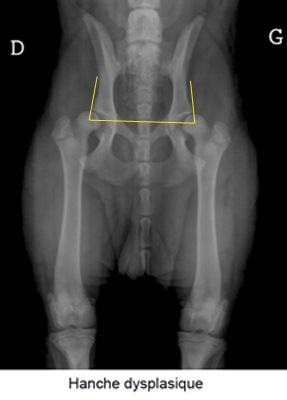
Coxofemoral dysplasia (hip dysplasia) is a common condition affecting mainly large breed dogs. This disease is due to abnormal development of the hip during the growth of the puppy. Heredity is a major factor in dysplasia. The parents may be nevertheless free from dysplasia, but they still have genes of the disease.

The first stage of hip dysplasia causes laxity of the hip joint. When the hip is supported, the hip becomes subluxated and as a result, the femoral head and the acetabulum (joint cavity) gradually deform as they flatten. Osteoarthritis appears on the joint and causes pain.

Sometimes the joint can be so altered that as a result of minor trauma it can dislocate. The animal then suddenly begins to limp. Most often, the condition is bilateral.

Generally, clinical signs of coxofemoral dysplasia can be detected around the age of 4 months, but diagnosis is more often made around 8 to 12 months. The main clinical signs are intolerance to exercise, gait, lameness, pain in hip extension, difficulty getting up after rest and atrophy of the muscles in the hindquarters. Some dogs do not show the first signs of dysplasia until around 2 years of age, or even when they are older after the onset of osteoarthritis.

The diagnosis of hip dysplasia is based on the combination of the animal's history, clinical signs present, orthopaedic and radiographic examinations. The operating decision is not made solely based on the radiographic examination, but takes into account the clinical context and the reality of the disability: "X-rays are not operated on".

The candidate for a triple osteotomy must have hip dysplasia, without osteoarthritis, a positive Ortalani sign (orthopaedic manoeuvre which makes it possible to highlight the laxity) with an angle of reduction lower than 40°, and especially the acetabulum must be retentive (the femoral head must enter the acetabulum in the radiographic position known as the frog). All candidates with a coxa-plana (flat cup), congenital hip dislocation or filled cup should be excluded from pat TPO therapy.

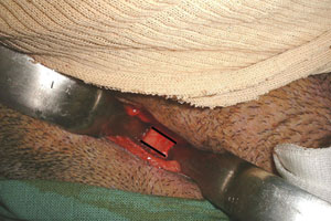
The procedure of the intervention

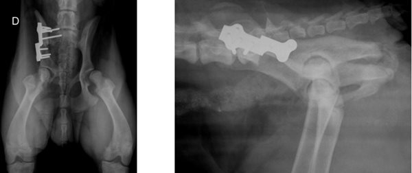
The principle of the operation is to make three cuts on the pelvic bone (triple osteotomy) to mobilise the acetabulum and tip it forward. This tilting effect increases the femoral head's dorsal overlap through the acetabulum and stabilizes the hip by neutralizing subluxation. 

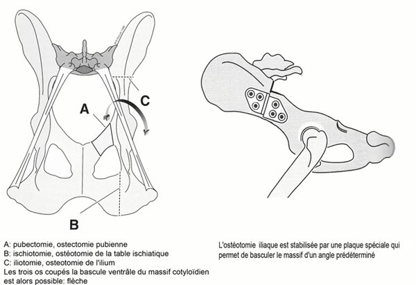
The intervention is carried out in three stages:

  1. Pubectomy: which consists of an inguinal approach to remove a small piece of the rising branch of the pubis
  2. The ischiotomy: consists of a caudal approach in cutting the ischiatic table in the sagittal plane.
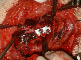
  3. The iliotomy: by the lateral approach of the ilium, a transversal section of the ilium is created which allows the acetabulum to be freed and tilted ventrally. This osteotomy is stabilized by a preformed osteosynthesis plate for triple pelvic osteotomy which allows giving exactly the desired angle of rotation.

1.étape 1 pubectomie 3.etape 3 iliotomie

After the operation, the patient must be strictly confined for 45 days, the resumption of pressure on the operated limb normally occurs in the first post-operative week, and lameness normally disappears in less than three weeks. Radiological normalization with progressive hip stabilization is performed over approximately three months to obtain optimal coverage.

Treatment Effectiveness

Triple pelvic osteotomy when indicated gives excellent results with normalization of gait and suppression of lameness.

Arthritic development is slowed and will normally be negligible and without functional consequences.

Most operated animals have a normal life without deficit on the operated hips, the operation can be bilateral.

This is the hip dysplasia correction procedure that is the most performed and favoured by most surgeons. It is a regulated surgical procedure for which the recoil and evolution of the implants currently guarantee the patient the best chances of an optimal quality of life if the surgical indication is correctly placed.

TPO is a complex procedure that requires advanced practice and knowledge of orthopaedic surgery.

Possible complications

- Regardless of the technique, the risk of anaesthesia accident, however low, remains non-zero.

- Surgical site infections are rare, and strict adherence to aseptic rules in orthopaedic surgery can prevent them.

- Too much activity during the healing period may lead to loosening of the implants, surgical resurfacing may be necessary but most of the time the bone segments position correctly and the acetabular cover remains satisfactory. After 6 weeks the healing is normally advanced enough for the implants to move.

- Trauma to the sciatic nerve may occur during the procedure, the neurological deficit is usually transient and resolves in less than three weeks. Exceptionally an irreversible lesion can occur, its consequence is catastrophic with a permanent loss of use of the limb.

- Trauma to the pelvic urethra can also occur, resulting in transient hematuria.

Nails and brooches

Nails and brooches

Nails and pins are one of the fundamental methods of fracture treatment. Spindles are also used as temporary stabilisation means in a large number of controlled surgical procedures. Although these osteosynthesis techniques can elegantly treat a large number of conditions, their relative stability in the rotation is their main deficiency, which is why they often have to be combined with other home stabilisation techniques: resin or complementary external fixator (known as the "Tied-in" technique).

Their great versatility always makes them a treatment of choice, particularly in juvenile skeletal orthopaedics where, when used in synergy with the image intensifier, they make it possible to effectively treat most fractures of the young person (see below). It is in this field that they find their main fields of indication.

Salter 2 complex fracture of the proximal tibial epiphysis with tuberous tear in a 5 month old puppy.

Diaphyseal fracture in a three-month-old puppy treated by fluoroscopically controlled closed-focus fasciculate nailing of the image intensifier.

Axial skeleton

Axial skeleton

It's the fractures that affect the spine. The first challenge for the veterinary orthopaedist with spinal traumatology is to be able to assess the neurological damage resulting from the fracture as safely as possible. In the vast majority of cases, lesions on nerve tissue are irreversible and prohibit surgical treatment to achieve satisfactory functional results. The preliminary neurological examination is therefore the key element of the surgical decision and its perspicacity. Normally fractures on the medullary territories cranially to the fifth lumbar vertebrae (L5) are of a very poor prognosis. Caudally at L6, the spinal cord stops to be prolonged by the nerves of the ponytail (root territories), these are much less sensitive to stretching and shearing and despite sometimes consequent displacements of fragments, the nervous deficits remain limited and make it possible to envisage reasonably a surgical intervention.


Ostéosynthèse Unilock du rachis chien

L5 body fracture with ventral lumbar displacement. Osteosynthesis by Unilock plate with implantation of screws in the root of thorny apophyses.

The preliminary neurological examination indicated a painful sensitivity preserved with the preservation of peripheral motor neurons.

Among the lumbar spine fractures, the L7 fracture with ventral sliding of S1 and sacrum is one of the most frequently encountered. Its surgical treatment, which involves reducing displacement and inserting trans-iliac pins, normally gives very good functional results.

Specific instrumentation

Specific instrumentation

Moteurs et ancillaires de pose

The placement of orthopaedic surgical implants requires the use of ancillary instruments and specific motors.

The Domitia veterinary clinic is equipped with dedicated air-powered orthopaedic motors. These motors consist of a handpiece to which accessories are fitted: mandrels, spindle pushers, bone saws or milling cutters... They are available in motors for large fragments (compact air drive "CAD" system), in motors for small fragments (Air Pen system), in an oscillating saw specific for osteotomies for levelling the tibial plateau (TPLO saw) and in a neurosurgical motor (Surgairtome 2). All these devices are tools specifically designed for orthopaedic surgery and are also widely used in human surgery. They are designed to be sterilised in humid heat (autoclave) and are the guarantee of a specific surgical exercise respecting the fundamental rules of prevention of nosocomial infections.

Scie Sécuros dédiée pour TPLO

Scie Sécuros dédiée pour TPLO

Moteur CAD Synthès pour gros fragments

Moteur CAD Synthès pour gros fragments

Brilliance Amplifier

The image intensifier is a radiology system with video control. It allows orthopaedic procedures to be performed in a closed home. Its main fields of application are nailing of the diaphysis and metaphyseal fractures with closed hearth, placement of external fixators, placement of implants in spine surgery. This is a fundamental element of modern orthopaedic surgery. It allows control over the position of the implants while reducing surgical trauma to its simplest expression.

Top of page
Surgeries

Practice information

Clinique vétérinaire Domitia

Back
  • Mon
    8h-12h 14h-19h
  • Tue
    8h-12h 14h-19h
  • Wed
    8h-12h 14h-19h
  • Thu
    8h-12h 14h-19h
  • Fri
    8h-12h 14h-19h
  • Sat
    8h-12h 14h-17h
  • Sun
    Closed
Back

Find us here:

44 rue Laurens Ravanel, 34500 Béziers
get directions with Google Maps