Principles
Principles
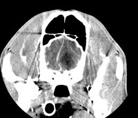
Computer-assisted axial tomography, otherwise known as X-ray scanning or simply scanning, is a medical imaging technique that allows radiographic images to be taken in anatomical sections perpendicular to the axis of the patient's body.
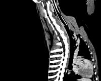
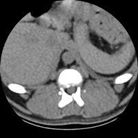
Axial Cuts - Lumbosacral Scanner of a dog - Sagital Reformatting. From the patient's native axial cuts, reconstruction software allows reformatting in all other planes (sagittal, coronal, or oblique). Finally, three-dimensional reconstructions can also be carried out with volume, surface, or endoscopic rendering.
Compared to conventional radiography, the scanner thus allows an overlay of anatomical structures and with a better contrast resolution.
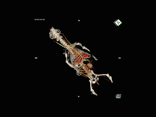
Full body 3D reconstructions with volume rendering
Examination Process
Examination Process
A general anesthetic is necessary for our four-legged patients: it limits the movements during the examination and allows reliable diagnostic images to be obtained: in fact, the slightest movement can compromise the quality and precision of the image. The animal must be left in the morning on an empty stomach and collected in the evening.
When the animal "sleeps", it is placed on the scanner bed in the appropriate position.
The scanner examination is quick and painless, the animal is exposed only to low doses of radiation.
The anaesthesia time is relatively short and the patient is monitored throughout the procedure.
The examination normally takes place in two phases, the first phase of image acquisition without preparation, followed by a second phase after injection of an intravenous iodinated contrast medium. The contrast agent is used to enhance vascular structures and certain tissues (especially tumours). It is the comparison of the two series of images that makes it possible to refine the tomodensitometric diagnosis.
Indications
Indications
The scanner gives the possibility to diagnose certain pathologies earlier and more precisely than with other imaging techniques (ultrasound or radiography). Like humans, the scanner allows veterinarians to view and examine parts of the body that are usually difficult to assess with conventional x-ray images.
The areas most commonly explored by CT scans in animals are the skull, spine, thorax, abdomen and certain joints such as the elbow or shoulder.
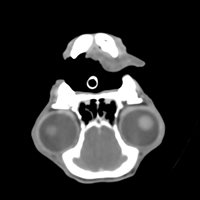
Nasal Cavities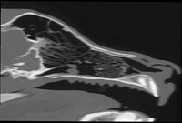
- Jetage and Chronic Rhinitis (runny nose)
- Epistaxis (nose bleeds)
- Deformities and pains of the muzzle
- Recurrent sneezing, reverse sneezing
- Suspicion of Aspergillosis, tumour or foreign body in the nasal cavities
- Sinusitis
- Periodontal diseases, Oro-Nasal fistulas (severe dental infections)
Facial mass
- Exophthalmos or asymmetry of the face
- Oral pain, difficulty opening or closing the mouth, trismus
- Temporal amyotrophy (muscle wasting)
- Claude Bernard Horner Syndrome
- Retrobulbar pathology
- Facial trauma
- Assessment of the extension of tumours on the face
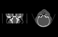
Tympanic bubbles
- Exploration of middle and internal ear infections
- Peripheral vestibular syndrome
- Facial paralysis
- Exploration of chronic otitis externa
- Suspicion of oropharyngeal polyps
- Exploration of tumours in the ENT sphere
Encephalon
- Exploration of Epileptic Seizures and Convulsive Syndromes
- Exploring unexplained sudden behavioural changes
- Exploration of alterations in the state of alertness
- Exploring Stroke
- Head injuries
- Exploration of pituitary Cushing's syndromes
- Amaurosis (loss of sight)
- Suspension of congenital diseases: Hydrocephaly, occipital dysplasia...
- Brain tumour suspension
Neckline
- Exploration of cervical fistulas and abscesses
- Exploration of salivary and thyroid glands
- Exploration of the cervical oesophagus
- Assessment of the extension of cervicofacial tumours and local-regional lymph nodes
Thorax
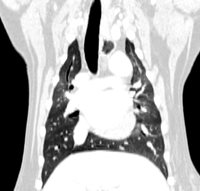
- Exploration of the thoracic masses
- Exploration of pleural effusions
- Research of foreign pulmonary bodies (epillets) and exploration of lung abscesses
- Exploration of pneumothorax
- Pulmonary extension assessment in oncology
- Exploration of congenital aortic arch malformations
Abdomen & Pelvis
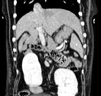
- Exploration of abdominal and pelvic masses
- Suspension of abdominal and pelvic foreign bodies
- Abdominal extension assessment in cancerology
- Exploration of portosystemic shunts by multiphase angioscan
- Exploration of abdominal organs: liver, spleen, pancreas, adrenals, kidneys, etc.
- Search for retroperitoneal foreign bodies
- Investigation of Cushing's syndromes
- Exploration of the urinary tree and search for ureteral ectopias
Axial Skeleton
- Suspicion of acute or chronic herniated discs
- Exploration of cervical vertebral instabilities and Wobbler's syndromes
- Traumatic assessment, exploration of vertebral fractures
- Suspicion of discospondylites (infection of discs and vertebral bodies)
- Exploration of Ponytail Syndrome
- Spinal and paraspinal tumour extension assessment
Skeleton Appendicular
- Exploration of elbow arthropathies: Dysplasia, Coronoid, Ancona, Osteochondritis, Incongruence
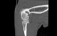
- Exploration of arthropathies of the shoulder: Tenosynovitis, Osteochondritis, Instabilities and lesion of the glenoid bead (arthroscanner)
- Exploration of the stifle (knee)
- Exploration of the carp and the Tarsus
- Bone tumour extension assessment
- Exploration of complex fractures: joints, acetabulum
- Regardless of the technique, the risk of anaesthesia accident, however low, remains non-zero.
- Given the injection of Iodine a renal check-up may be necessary.
- For older animals, a complete pre-anaesthetic check-up may be essential.
- Exceptionally, a severe allergic reaction may occur with the contrast material.