Neurology

Neurology consultations are dedicated to animals presenting symptoms suggestive of a disease of the nervous system or muscles:


  • convulsive seizures,
  • paralysis of one or more limbs,
  • compulsive walking,
  • going round in circles,
  • deafness,
  • loss of vision of nervous origin,
  • weakness…
  • Atlanto-Axial Instability
  • Laminectomy: Disc Herniation
  • Cauda Equina : Horsedick

Atlanto-Axial Instability

Herniated Disc

- Herniated discs are a very common pathology in dogs and rarer in cats.

- They cause compression of nerve tissue resulting in pain and musculoskeletal disorders that can lead to paralysis.

- Their treatment most often involves surgery with good results in 90% of cases operated on.

Introduction

Herniated disks are caused by breaking into the spinal canal of part of the intervertebral disc. This intrusion causes compression of nerve tissue.

Herniated discs may occur at the cervical, thoracolumbar or lumbar levels. Their highest frequency is at the thoracolumbar level.

The consequences of the medullary compression are pain and especially locomotor disorders, that can go as far as paralysis.

Atlanto-Axial Instability

It is a malformation affecting the first two cervical vertebrae (Atlas "C1" and Axis "C2").

- It is a pathology affecting miniature and Toy breeds such as: Yorkshire, Poodles, Pekingese, Shih-Tzu.

- The anomaly of the vertebrae leads to major instability which consequences are cervical pain and neurological locomotor disorders.

- Treatment is surgical and involves stabilising the vertebrae. The results are good in 90% of cases.

Introduction

Atlantic-axial instability is a malformation of the first two cervical vertebrae (Atlas and Axis) that causes excessive flexion of the vertebrae and dorsal displacement of the second cervical vertebrae (Axis). This displacement causes compression of the C2 medullary segment.

Normal anatomy of the occipital region and the atlanto-axial joint (C1-C2)

The disease mainly affects toy breeds, it is often the consequence of a malformation of the second cervical vertebrae (agenesis of the odontoid process). As a result of a small cervical trauma, the intervertebral ligament ruptures and C2 subluxation occurs. Subluxation then leads to compression of nerve tissue in the spinal canal, resulting in pain and motor neurological disorders (ataxia with para or tetraparesis or even paralysis).

Odontoid process agenesis (Axis tooth) and subluxation with Atlanto-Axial instability

In certain major cervical traumas, a fracture of the odontoid process may occur, with the same consequences as the absence of a tooth on the axis.

Fracture de la dent de l'axis et Subluxations Atlanto-Axiale

The procedure of the intervention

The operation is done under general anaesthesia, it consists in stabilising the first two cervical vertebrae. There are techniques by dorsal or ventral approach. The technique which currently has the favour of a large number of surgeons is arthrodesis by the ventral way.

Diagnosis is made by X-rays of the cervical region and possibly by scanner or myelography. Cervical spine manipulation should be done very carefully if atlanto-axial instability is suspected in order to avoid aggravating medullary compression.


Myelographic images show the agenesis of the axis tooth and medular compression.

The operation consists, after the approach of the ventral face of C1and C2 to abrade the articular surfaces of the atlanto-axial joint and to screw between them the vertebrae in order to carry out an interbody arthrodesis. A cancellous bone graft is put in place to help consolidate the arthrodesis.

The aspect of the Atlanto-Axial screwing at the end of the operation

The use of an intraoperative image intensifier allows a secure positioning of the implants. The main risk of this operation is a vicious positioning of the screws with a significant risk of spinal cord injury. Fluoroscopic control makes it possible to adjust the screws in their installation corridor with a precision that visual assessment alone cannot achieve.

Post-operative radiography of Atlanto-Axial stabilization


Post-Operative and Results

A neck brace is left around the patient's neck for two to three weeks. Meals must be served high during this period. Exercise and outings must be strictly controlled for 45 days and the collar must be replaced with a harness.

Short, medium and long term results are excellent in 90% of cases. The pain disappears in the days following the operation and the locomotor disorders within a few weeks.

The atlanto-axial arthrodesis remains a delicate neurosurgical operation, the constraints of placement of implants impose its realisation by experienced surgeons practising within correctly equipped establishments.

Possible complications

- Regardless of the technique, the risk of anaesthesia accident, however low, remains non-zero.

- Surgical site infections are rare and generally benign.

- A serous collection can be built up on the surgical site without functional consequences.

- The greatest danger of the operation is the rupture of the medullary canal by an implant and irreversible trauma to the marrow. The consequences are catastrophic and irreversible. This risk can be effectively reduced by using a brightness amplifier.

Laminectomy: Disc Herniation

Laminectomy: Disc Herniation

Axial cuts

Scanner of an Extrusive T13L1 Herniated Disc in a Dog & sagittal and coronal reformatting

Laminectomy and Hemilaminectomy

After general anaesthesia, the animal is placed in a ventral position.

The principle is to approach the spine dorsally or dorsolateral. Part (hemilaminectomy) or all (laminectomy) of the vertebral dorsal blade is removed with a neurosurgical bur and special rodents. It is thus practised a window in the vertebra which allows at the same time to decompress the nervous tissues and to reach the herniated discal material which can be removed.

After a laminectomy, recovery times are variable, depending on the initial stage of the lesions. The higher the stage of the lesion, the longer recovery times vary from a few days to several months. They also depend on the disc herniation stage, the lower lumbar hernias in root territory recover much faster than the thoracolumbar hernias.

Stabilisation of the cervical spine by locking screws and titanium intersomatic plates.

tabilisation du rachis cervical hernie cervicale

Prognosis

The key element of prognosis is the initial neurological stage.

  • Stage 1 to 3 herniated discs have recovery rates greater than 90% with surgical treatment.
  • Stage 4 herniated discs have recovery rates between 85% and 90% depending on the authors.
  • stage 5 hernias are much more difficult to treat. in individuals operated immediately after the onset of paralysis and deep painful loss of sensation (less than six hours) can expect recovery rates around 70%. For animals evolving for more than six hours, the results do not meet consensus, it is generally accepted that between 6 and 12 hours 50% good results can be expected and that rates fall below 30% beyond 12 hours (or 24 hours depending on the authors).

Possible complications

- Regardless of the technique, the risk of anaesthesia accident, however low, remains non-zero.

- Surgical site infections are rare and generally benign.

- A serous collection can be built up on the surgical site without functional consequences.

- There is often a worsening of the patient's neurological status after the procedure. This stapling is normally transient.

- Despite surgical treatment and with correct initial neurological stages, some patients do not recover motor skills. Hernias of the chronic high disc protrusion type are those for which the operation is the most delicate.

Cauda Equina : Horsedick

Cauda Equina : Horsedick (Ponytail Syndrome)

- It is a pathology affecting the most caudal part of the lumbar spine and is comparable to the pathology of the sciatic nerve in men.

- It leads to a compression of the roots of the sciatic nerve which causes pain and locomotor disorders.

- The causes are varied: a herniated disc, narrow channel syndrome, vertebral instability, fracture, infection of the intervertebral disc, tumour...

- The evolution of ponytail syndrome (PTS) is normally chronic but acute forms may also exist.

- The SQC affects rather large breed dogs (German Shepherd, Labrador...) but toy dogs or cats can also be affected.

- Surgical treatment is effective if patients are operated on as early as possible before degenerative nerve damage occurs.

Introduction

Ponytail syndrome is a pathology of the lumbosacral junction and the caudal lumbar spine, it concerns regions of the spinal column after the end of the spinal cord for which only the nerve roots of the sciatic and sacral nerves exist, it is a pathology called the root stage.

It normally reaches animals of middle to advanced age and evolves in a chronic mode with a progressive degradation of motor skills and sensitivity in the territories of the sciatic nerve. However, early progression in the case of congenital malformations or acute presentations in the case of trauma or herniated discs is also possible. SQC is always accompanied by pain on the lumbosacral level, which is largely responsible for the animal's discomfort.

Anatomical elements and symptomatology

Compression concerns so-called root areas of the spine. Because of a growth differential during development, the spinal cord grows less than the spine, so there is a gap between the spinal and spinal segments. This shift is noticeable from the second lumbar vertebra (L2) and has as a consequence the end of the marrow (medullary cone) opposite the 5th lumbar vertebra. On the other hand, the spinal nerves continue to leave the vertebral canal through their respective foramens at the level of their corresponding vertebral segment. Thus, beyond L5-L6, only the nerves and the terminal part of the meninges (dural cone) remain, which by their filament aspect constitute the ponytail.

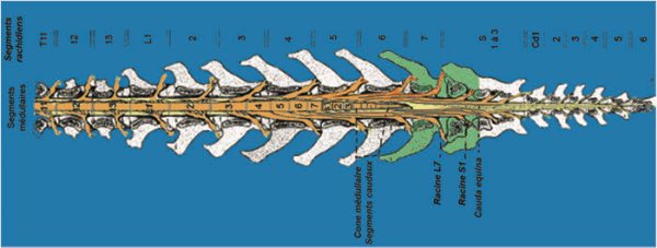
Cross-section view in the frontal plane of the terminal spine: notice the medullar cone facing L5 and L6 and beyond the medullar canal occupied only by nerves (oranges) stretched like the hairs of a ponytail.

Schematically, after leaving the spinal canal, the nerve bundles group together to form the sciatic nerve (roots L6 to S1) and the pelvic and shameful nerves (roots S1-S3). As a result, the intra-channel compressions after the sixth lumbar vertebra will result in nervous disorders on the sciatic nerves and pelvic nerves. Carnivore ponytail syndrome is equivalent in some ways to sciatica in humans.

The causes of compression of the ponytail are numerous, the lumbosacral junction (L7-S1) is a hinge zone of the spine with the connection of the spinal column to the pelvis via the sacrum. It is an area subject to significant mechanical stresses in bending and extension.

Congenital stenoses of the vertebral canal: the vertebral canal is too narrow and compresses the nervous tissue, these stenoses can be associated with malformations of the spine: the sacralization of the last lumbar vertebra, non-union of the sacral vertebrae... Congenital stenosis may have pathological consequences only late with the ageing of ligament structures and their thickening.

  • Spinal instabilities: spondylolisthesis L7S1 for example.
  • Herniated discs posterior to the end of the medullary cone.
  • Intervertebral disc infection: spondylodiscitis, lumbosacral discospondylitis.
  • Neoplasms of the spine or nerve tissue.

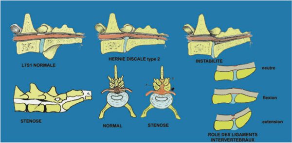
Major Non-Tumor Causes of Ponytail Syndrome

The symptoms are varied and their progressive appearance, most of the time the ponytail syndrome evolves on a chronic mode with a progressive degradation of the patient, exceptionally the evolution can be made on an acute mode in particular in case of an acute herniated disc or vertebral fracture/dislocation. The progression is often insidious.

  • Pain at the lumbosacral junction is normally constant in ponytail syndrome and for a long time the only symptom present, this pain should not be confused with that which may be caused by coxofemoral dysplasia.
  • A proprioceptive deficit on the hind legs results in excessive wear of the claws and scraping of the ground.
  • Posterior ataxia.
  • Muscular cast iron on the hind limbs.
  • Decreased sciatic reflexes (withdrawal reflex and possibly patellar pseudo-hyperreflexia).
  • Atony of the tail.
  • Urinary and/or faecal incontinence in the most advanced cases.

The procedure of the intervention

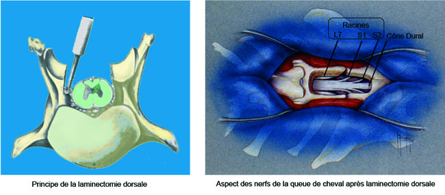
The operation is done under general anaesthesia, it consists in decompressing the nerve roots.

The most common procedure is a dorsal laminectomy of the L7 and S1 vertebrae (recalibration). This laminectomy can be extended to intervertebral foramens and joint apophyses to extend decompression to the roots in their foraminal path. Except in the case of a vertebral fracture, no additional stabilisation is generally performed in veterinary surgery. A discectomy (removal of the intervertebral disc) is also performed during the procedure in most cases.

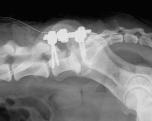
In certain cases, notably when a very large laminectomy with extension to the intervertebral joints must be performed in order to treat intervertebral foramen stenosis or, in case of the existence of a very important spondylolisthesis, a complementary stabilisation of the spine must be performed. This is carried out by the installation of pedicle screw systems and union bars or locked titanium plates. These implants block the spinal movements and allow a faster recovery by facilitating the fusion of the vertebral bodies.

Lousewort screwing by Axon Synthes system after extended L7S1 laminectomy
for herniated disc with foraminal stenosis


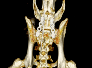
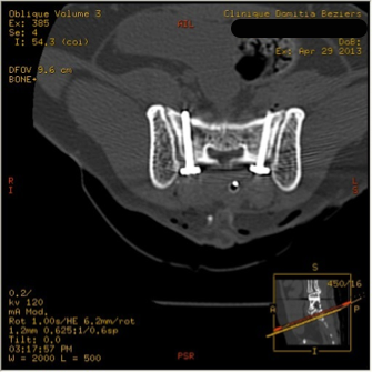
Lumbosacral stabilisation by pedicle screw and Unilock Synthès plates in a cat


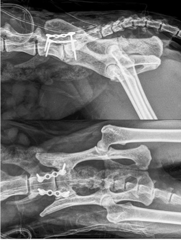
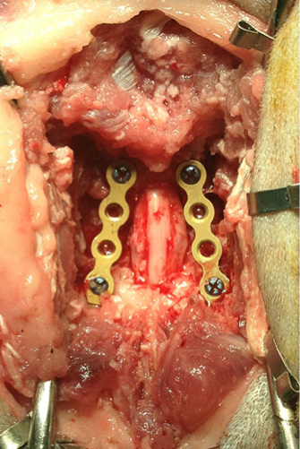
Stabilisation of lumbosacral junction after laminectomy and bilateral foraminectomy in a cat by pedicle screws and Unilock Synthès plates

vissage pédiculaire chat scanner 3DVR 

vissage pédiculaire vissage scanner axial

Alternatively to dorsal laminectomy, an intervertebral arthrodesis in flexion can be performed, this technique is very rarely practised in France where the majority of surgeons prefer dorsal decompression.

Treatment Effectiveness

For animals operated early the effectiveness of the treatment is excellent with 90% good results. The pain is eliminated immediately and the locomotor disorders disappear within a few weeks. Patients regain the comfort of living and walking perimeter they had lost.

In advanced cases in which neurological degeneration is already present, the improvement is noticeable; however, some permanent nerve lesions cannot be recovered, but the evolution of the degenerative process is stopped.

From a postoperative point of view, at the root level, there is little or no motor impairment in the immediate postoperative period and patients are usually ambulatory the day after the operation.

Possible complications

- Regardless of the technique, the risk of accident to anaesthesia, however low, remains non-zero.

- Surgical site infections are rare and generally benign.

- A serous collection can be built up on the surgical site without functional consequences.

- Exceptionally a trauma of the nerve roots can intervene, this one can have catastrophic consequences.

Top of page
Surgeries

Practice information

Clinique vétérinaire Domitia

Back
  • Mon
    8h-12h 14h-19h
  • Tue
    8h-12h 14h-19h
  • Wed
    8h-12h 14h-19h
  • Thu
    8h-12h 14h-19h
  • Fri
    8h-12h 14h-19h
  • Sat
    8h-12h 14h-17h
  • Sun
    Closed
Back

Find us here:

44 rue Laurens Ravanel, 34500 Béziers
get directions with Google Maps